Protocol Overview
This diagram shows an overview of the protocol presented here:
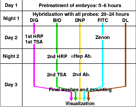
- Currently, we are working with four commonly used probe labels,
- Digoxigenin (DIG)
- Biotin (BIO)
- Dinitrophenyl (DNP)
- Fluorescein (FITC)
and using standard immunofluorescence techniques to detect these labels. For each label, there are multiple ways to achieve the fluorescent visualization, but for the sake of simplicity, the diagram shows only one way for each label.
- Sequential tyramide signal amplification (TSA) reactions using horseradish peroxidase (HRP) conjugates give very sensitive and reliable two-color fluorescence.
- Fluorescent secondary antibodies (2nd Ab.) can be used to label primary antibodies (anti-Hap Ab.) against the probe labels, or 'haptens'.
- 'Zenon' refers to a method of making fluorescently labeled antibody complexes, which are then incubated with embryos. Not only are you freed from maintaining species separation with your antibodies, but also you can visualize the probes with only one incubation and washing step.
- We are also experimenting with directly labeled (DL) probes, into which fluorescent dye molecules of the 'Alexa' series have been incorporated. These probes can be visualized on embryos immediately after hybridization.
- For more on sequential tyramide reactions, zenon antibody labeling, and direct label probes, see Alternative Methods for Fluorescent Detection.
- You can combine any and all of these methods to get a multi-color fluorescent stain, providing great flexibility in the design of experiments. A more detailed discussion of them can be found here under Conclusions and Future Problems.
Next: Embryo Fixation
Previous: Embryo Gallery
Back to Index